 Genes
Genes
 Phenotypes & Mutant Alleles
 Human–Mouse: Disease Connection
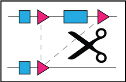 Recombinase (cre)
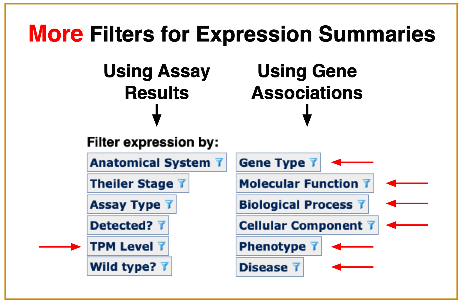
 Function
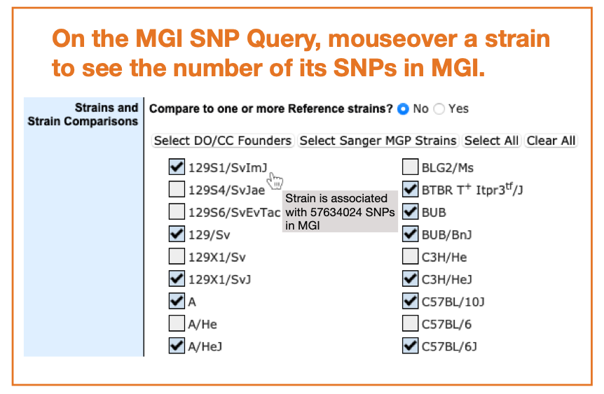
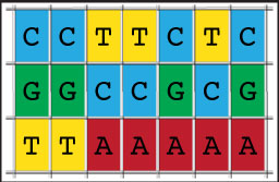 Strains, SNPs & Polymorphisms
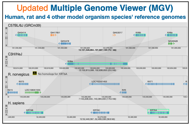
 Vertebrate Homology
 Mouse Models of Human Cancer
 Batch Data and Analysis Tools
 Nomenclature

The International Mouse Phenotyping Consortium project is systematically phenotyping knockout mice from the mutant ES cells produced by the International Mouse Knockout Consortium. Data will be integrated into MGI as available.
Mouse Genome Database (MGD), Gene Expression Database (GXD), Mouse Models of Human Cancer database (MMHCdb) (formerly Mouse Tumor Biology (MTB)), Gene Ontology (GO) |
||
|
Citing These Resources Funding Information Warranty Disclaimer, Privacy Notice, Licensing, & Copyright Send questions and comments to User Support. |
last database update 04/23/2024 MGI 6.23 |
|
|
|
||
 Analysis Tools