|
|
|
|
|
|
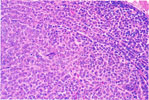
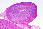
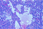
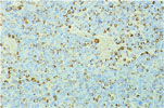
| 121. Adrenocortical Adenoma (Type B Solid Cell) |
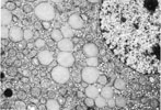
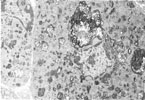
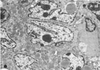
122.
Electron Micrograph (TEM) -Adrenal Cortical Epithelial Cells |
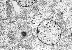
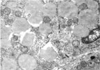
123.
Electron Micrograph (TEM) -Adrenal Medullary Epithelial Cells |
124.
Electron Micrograph -Ceroid Pigment -Adrenal |
125.
Electron Micrograph -Ceroid Pigment -Adrenal |
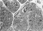
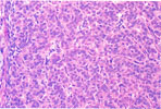
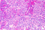
126. Adrenocortical Carcinoma (Type A Cell) |
|
|
|
|

|
|
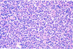
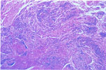
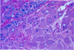
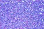
| 127. Adrenocortical Carcinoma (Type B Cell) |
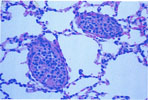
128.
Pulmonary Metastasis -Adrenocortical Carcinoma (Type B Cell) |
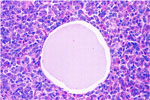
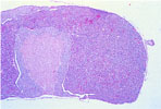
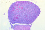
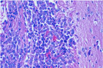
129. Diffuse Adrenal Medullary Hyperplasia | 130. Pheochromocytoma |
131.
Pulmonary Metastasis -Pheochromocytoma |
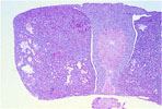
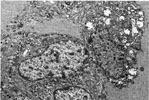
132.
Ganglioneuroma -Adrenal |
|
|
|
|
|
|
|
133.
Ganglioneuroma -Adrenal |
134.
Electron Micrograph -Adrenocortical Type A Carcinoma (Lipid Droplets) |
135.
Electron Micrograph -Adrenocortical Type A Carcinoma (Desmosome) |
136. Pituitary Cyst |
137.
Cystoid Degeneration -Pituitary |
138.
Cystoid Degeneration -Pituitary |
|
|
|
|
|
|
|
139.
Focal Hyperplasia -Pituitary |
140.
Focal Hyperplasia -Pituitary |
141. Pituitary Adenoma |
142.
Pituitary Adenoma -Prolactin |
143. Pituitary Carcinoma Invading Brain |
144.
Electron Micrograph -Pituitary Cystoid Degeneration |