|
|
|

|
|
|
|
49.
Pinworms -Colon |
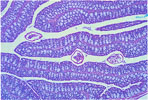
50.
Small Adenoma (Polyp) -Small Intestine |
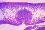
51.
Large Adenoma (Polyp) -Large Intestine |
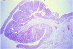
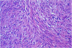
52. Intestinal Adenocarcinoma | 53. Intestinal Leiomyosarcoma |
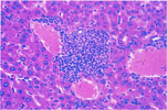
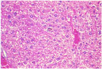
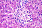
54.
Granulopoietic Hyperplasia -Liver |
|
|
|
|
|
|
|
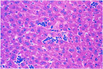
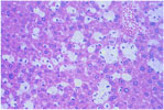
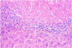
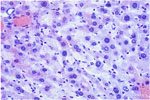
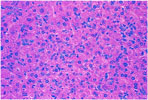
55.
Erythropoietic Hyperplasia -Liver |
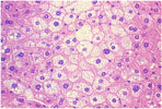
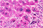
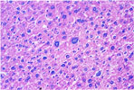
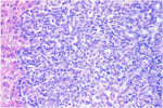
56.
Fatty Metamorphosis -Liver (H & E) |
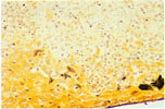
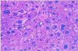
57.
Fatty Metamorphosis -Liver |
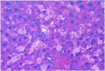
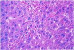
58.
Lipid Storage Disease -Liver |
59.
Hemosiderosis -Kupffer Cells -Liver (H & E) |
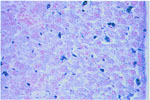
60.
Hemosiderosis -Kupffer Cells -Liver (Prussian Blue) |
|
|
|
|
|
|
|
61.
Ceroid Pigment -Liver |
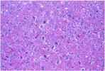
62.
Coagulative Necrosis -Liver |
63.
Giant Cells -Mouse Hepatitis Virus Infection -Liver |
64.
Tyzzer's Disease -Liver (Warthin-Starry Stain) |
65.
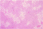
Amyloidosis -Liver (H & E) |
66.
Amyloidosis -Liver (Congo Red) |
|
|
|
|
|
|
|
67.
Karyomegaly and Cytomegaly -Liver |
68.
Intranuclear Inclusion -Hepatocyte |
69.
Intracytoplasmic Inclusions -Hepatocytes |
70.
Bile Duct Hyperplasia -Liver |
71.
Oval Cell Hyperplasia -Liver |
72.
Cholangiofibrosis (Adenofibrosis) -Liver |