|
|
|
|
|
|
|
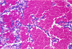
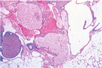
217.
Angiectasis -Ovary |
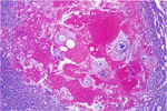
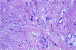
218.
Ectopic Pregnancy -Ovary |
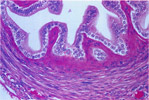
219.
Fibrosis -Oviduct |
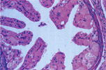
220.
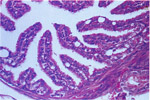
Hyaline Change -Oviduct |
221.
Vacuolization -Oviduct |
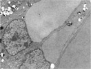
222. Electron Micrograph of Oviduct Hyaline Change |
|
|
|
|
|
|
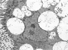
| 223. Electron Micrograph of Oviduct Vacuolization |
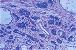
224.
Tubular Adenoma (Tubular Mesothelioma) -Ovary |
225.
Tubular Adenoma -Ovary |
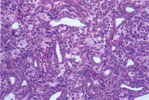
226.
Tubular Carcinoma -Ovary |
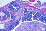
227.
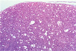
Serous Cystadenoma -Ovary |
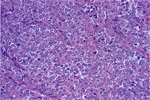
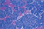
228.
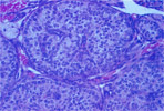
Dysgerminoma -Ovary |
|
|
|
|
|
|
|
229.
Teratoma -Ovary |
230.
Teratoma -Ovary |
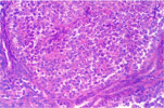
231.
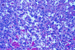
Granulosa Cell Tumor -Ovary |
232.
Cal-Exner Body -Ovarian Granulosa Cell Tumor |
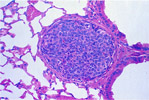
233.
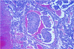
Pulmonary Metastasis -Ovarian Granulosa Cell Tumor |
234.
Thecoma -Ovary |
|
|
|
|
|
|
|
235.
Luteinized Thecoma -Ovary |
236.
Luteoma -Ovary |
237.
Sertoli Cell Tumor -Ovary |
238.
Hemangioma -Ovary |
239.
Hemangiosarcoma -Ovary |
240.
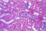
Flattened Bowman's Parietal Epithelium -Female Mouse |