|
|
|
|
|
|
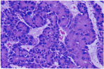
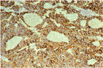
| 25. Parotid Adenocarcinoma |
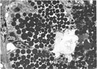
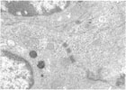
26.
Electron Micrograph -Male Submaxillary Gland -Granules |
27.
Electron Micrograph -Female Submaxillary Gland |
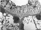
28.
Electron Micrograph -Myoepithelioma -Fibrils |
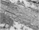
29.
Electron Micrograph -Myoepithelioma -Desmosomes |
30.
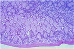
Pancreatic Acinar Atrophy with Fatty Replacement |
|
|
|
|
|
|
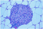
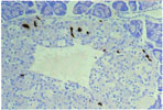
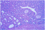
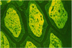
| 31. Pancreatic Islet Cell Hyperplasia |
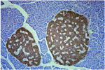
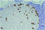
32.
Immunoperoxidase -Insulin -Islet Hyperplasia |
33.
Immunoperoxidase -Somatostatin -Islet Hyperplasia |
34.
Immunoperoxidase -Glucagon -Islet Hyperplasia |
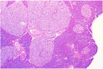
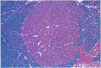
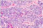
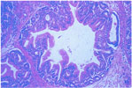
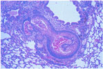
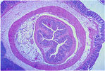
35. Islet Cell Adenoma | 36. Islet Cell Carcinoma |
|
|
|
|
|
|
|
37.
Islet Cell Carcinoma -Somatostatin |
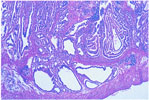
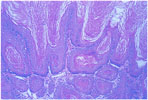
38. Gastric Glandular Hyperplasia |
39.
Gastric Adenoma -Glandular Stomach |
40. Gastric Adenocarcinoma |
41.
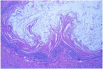
Squamous Hyperplasia -Forestomach |
42.
Squamous Cell Papilloma -Forestomach |
|
|
|
|
|
|
|
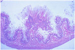
43.
Squamous Cell Papilloma -Forestomach |
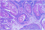
44.
Squamous Cell Carcinoma -Forestomach |
45.
Pulmonary Metastasis -Gastric Squamous Cell Carcinoma |
46. Intestinal Amyloidosis (H & E) | 47. Intestinal Amyloidosis (Thioflavin T) | 48. Intestinal Intussusception |