|
|
|
|
|
|
|
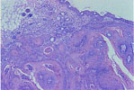
313.
Squamous Cell Carcinoma Arising in a Papilloma -Skin |
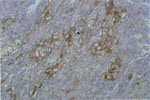
314.
Squamous Cell Carcinoma -Keratin |
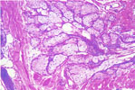
315.
Sebaceous Gland Adenoma -Skin |
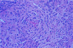
316.
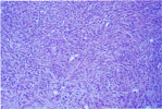
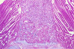
Spindle Cell Sarcoma -Subcutis |
317.
Hemangiopericytoma -Subcutis |
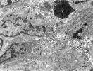
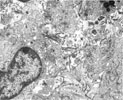
318.
Electron Micrograph -Fibrosarcoma -Collagen Production |
|
|
|
|
|
|
|
319.
Electron Micrograph -Fibrosarcoma -Collagen Production |
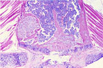
320.
Early Fibro-osseus Lesion -Bone |
321.
Advanced Fibro-osseus Lesion -Bone |
322.
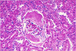
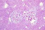
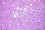
Osseous Metaplasia -Kidney |
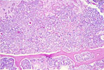
323. Osteosarcoma | 324. Osteosarcoma |
|
|
|
|
|
|
|
325.
Hepatic Metastasis -Osteosarcoma |
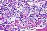
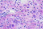
326. Chondrosarcoma |
327.
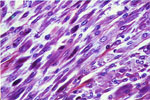
Rhabdomyosarcoma -Skeletal Muscle |
328.
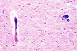
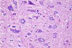
Cerebral Calcification -Thalamus |
329.
Inclusion Cyst -Spinal Cord |
330.
Inclusion Cyst -Brain |
|
|
|
|
|
|
|
331.
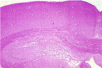
Hydrocephalus -Lateral Ventricle |
332.
Lipid Storage Disease -Decrease in Purkinje Cells -Cerebellum |
333.
Lipid Storage Disease -Cerebrum |
334.
Vacuolization of White Matter -Brain |
335. Cerebral Infarct |
336.
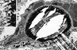
Electron Micrograph -Cerebral Thalamic Calcification |