|
|
|
|
|
|
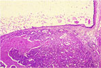
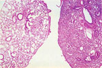
| 97. Gall Bladder Carcinoma |
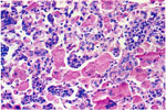
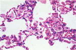
98.
Eosinophilic Crystals -Pulmonary Alveolar Macrophages |
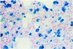
99.
Chronic Passive Congestion (Prussian Blue Stain) -Lung |
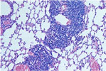
100.
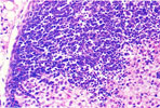
Perivascular and Peribronchial Lymphoid Aggregates -Lung |
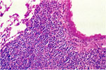
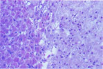
101. Pulmonary Atelectasis Adjacent to Normal Lung | 102. Pulmonary Alveolar Macrophage Accumulations |
|
|
|
|
|
|
|
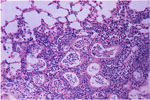
103.
Mycoplasma pulmonis Infection -Lungs |
104.
Chronic Sendai Virus Infection -Lungs |
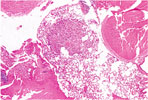
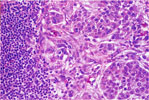
105. Pulmonary Alveolar Cell Adenoma | 106. Pulmonary Alveolar Cell Adenoma |
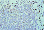
107.
Adenoma -Pulmonary Surfactant Apoprotein -Lung |
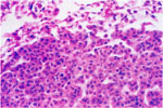
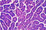
108.
Papillary Adenocarcinoma -Lung |
|
|
|
|
|
|
|
109.
Lymph Node Metastasis of Pulmonary Adenocarcinoma -Lung |
110.
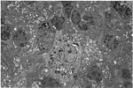
Electron Micrograph -Pulmonary Alveolar Type II Adenoma |
111.
Electron Micrograph -Pulmonary Alveolar Type II Adenoma |
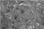
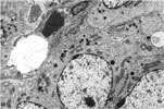
112.
Electron Micrograph -Papillary Adenocarcinoma -Lung |
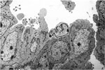
113.
Electron Micrograph -Papillary Adenocarcinoma -Lung |
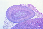
114.
X-Zone -Adrenal Cortex |
|
|
|
|
|
|
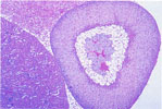
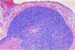
| 115. Adrenocortical Vacuolization (X-Zone) | 116. Accessory Adrenocortical Nodule | 117. Subcapsular Adrenocortical Spindle Cell Hyperplasia |
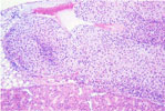
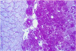
118.
Ceroid Pigment -Macrophages -Zona reticularis -Adrenal (H & E) |
119.
Ceroid Pigment -Adrenal (PAS) |
120. Adrenocortical Adenoma (Type A Spindle Cell) |
| Back |