|
|
|
|
|
|
|
1.
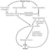
The Potential Histopathogenesis of Cancer in Mice |
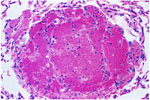
2.
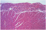
Epicardial Mineralization (H & E) |
3.
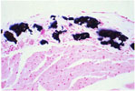
Epicardial Mineralization (von Kossa) |
4.
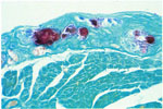
Epicardial Mineralization (Alizarin Red) |
5.
Myocardial Mineralization |
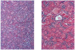
6.
Atrial Thrombosis -Left Atrium |
|
|
|
|
|
|
|
7.
Atrial Thrombosis -Cartilaginous Metaplasia |
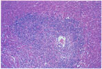
8.
Medial Vascular Hypertrophy |
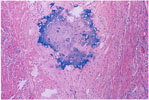
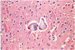
9.
Medial Mineralization -Thalamus |
10.
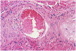
Coronary Atherosclerosis |
11.
Coronary Atherosclerosis |
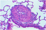
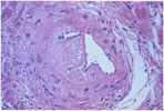
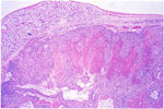
12.
Coronary Polyarteritis |
|
|
|
|
|
|
|
13.
Ovarian Angiectasis |
14.
Hepatic Angiectasis |
15.
Hemangioma -Uterus |
16.
Hemangiosarcoma -Liver |
17.
Pulmonary Metastasis -Hepatic Hemangiosarcoma |
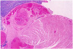
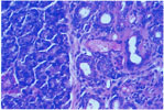
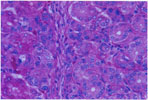
18.
Female and Male Submaxillary Salivary Glands |
|
|
|
|
|
|
|
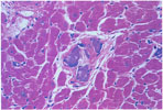
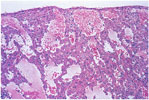
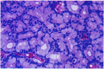
19.
Lobular Atrophy -Salivary Gland |
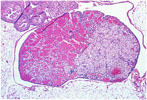
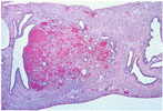
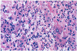
20.
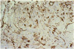
Salivary Gland Myoepithelioma |
21.
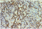
Myoepithelioma -Keratin |
22.
Myoepithelioma Intermediate Filaments -Vimentin |
23.
Adenoma -Serous Cells -Submaxillary Salivary Gland |
24.
Adenoma -Mucous Cells -Submaxillary Salivary Gland |