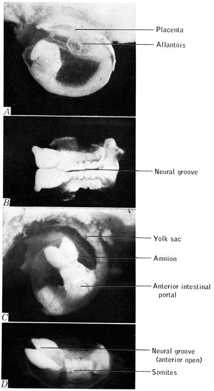
Figure 12-26. Photographs (x 22.5) of mouse embryos. A. Lateral view of 7-day 18-hour, 6-somite embryo, with decidua and most of yolk sac dissected. B. Dorsal view of same embryo, amnion also dissected. C. 10-somite embryo, age 9 to 9½ days. Slightly retouched. Embryo from inbred stock. D. Same embryo as C, dorsal view, amnion removed.