Phenotypes associated with this allele
|
|
| Find Mice |
Using the International Mouse Strain Resource (IMSR)
Mouse lines carrying:
Tg(Myhca-cre)1Abel mutation
(0 available)
Tmsb4xtm1.2Chen mutation
(0 available);
any
Tmsb4x mutation
(11 available)
|
|
|
normal phenotype
 |
• mutant mice are viable to adulthood
|
|
|
| Find Mice |
Using the International Mouse Strain Resource (IMSR)
Mouse lines carrying:
Bag3tm1.1Chen mutation
(0 available);
any
Bag3 mutation
(31 available)
Bag3tm1c(EUCOMM)Hmgu mutation
(1 available);
any
Bag3 mutation
(31 available)
Tg(Myhca-cre)1Abel mutation
(0 available)
|
|
|
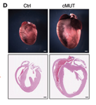
Cardiac enlargement in Bag3tm1c(EUCOMM)Hmgu/Bag3tm1.1Chen Tg(Myhca-cre)1Abel/0 mice at 6 months of age
cardiovascular system
|
|
• heart weight to body weight and heart weight to tibia length ratios are increased at 5 months of age
|
|
|
• diminished systolic function
|
|
|
• diminished systolic function with mice showing reduced fractional shortening, increased left ventricle internal diameter at end diastole and increased left ventricle internal diameter at end systole
|
homeostasis/metabolism
cellular
|
|
• hearts exhibit impaired autophagic flux
|
muscle
|
|
• diminished systolic function
|
growth/size/body
|
|
• heart weight to body weight and heart weight to tibia length ratios are increased at 5 months of age
|
|
|
| Find Mice |
Using the International Mouse Strain Resource (IMSR)
Mouse lines carrying:
Slc2a4tm1Abel mutation
(0 available);
any
Slc2a4 mutation
(35 available)
Tg(Myhca-cre)1Abel mutation
(0 available)
|
|
|
cardiovascular system
|
|
• during normal perfusion, glycogen content is 54% higher in fed mutants than in fed wild-type, however no difference in myocardial glycogen in fasted mice
|
|
|
• during normal perfusion, hearts from fed homozygotes show higher concentrations of phosphocreatine
|
|
|
• during ischemia, hearts from fasted homozygotes exhibit depressed glucose utilization
|
|
|
• during normal perfusion, hearts from fed homozygotes show a 3-fold increase in glucose uptake, while fasted hearts show identical levels of basal glucose uptake as wild-type
|
|
|
• fasting results in a 20% decrease in left ventricular systolic pressure, however heart rates and coronary flow rates are unaffected by fasting
|
|
|
• hearts exhibit delayed recovery after low-flow ischemia
• hearts of fasted mutants exhibit decreased lactate production (75%) indicating depressed glucose utilization during ischemia and develop irreversible systolic and diastolic dysfunction as indicated by an increase in left ventricular end-diastolic pressure that remains elevated on reperfusion and a left ventricular developed pressure that does not recovery fully, as well as impaired ability to generate ATP during ischemia (accelerated depletion of ATP and decreased levels of ATP recovery on reperfusion)
• hearts of fed mutants exhibit normal left ventricular pressure during ischemia, however recovery of the left ventricular developed pressure is delayed during reperfusion and hearts exhibit a decrease (28%) in lactate production during ischemia
|
homeostasis/metabolism
muscle
|
|
• during normal perfusion, glycogen content is 54% higher in fed mutants than in fed wild-type, however no difference in myocardial glycogen in fasted mice
|
|
|
• during ischemia, hearts from fasted homozygotes exhibit depressed glucose utilization
|
|
|
• during normal perfusion, hearts from fed homozygotes show a 3-fold increase in glucose uptake, while fasted hearts show identical levels of basal glucose uptake as wild-type
|
cellular
|
|
• during ischemia, hearts from fasted homozygotes exhibit depressed glucose utilization
|
|
|
• during normal perfusion, hearts from fed homozygotes show a 3-fold increase in glucose uptake, while fasted hearts show identical levels of basal glucose uptake as wild-type
|
|
|
| Find Mice |
Using the International Mouse Strain Resource (IMSR)
Mouse lines carrying:
Slc2a4tm1Abel mutation
(0 available);
any
Slc2a4 mutation
(35 available)
Tg(Myhca-cre)1Abel mutation
(0 available)
|
|
|
cardiovascular system
|
|
• cross sectional area of myocytes was greater
• gross morphology of the heart was normal however
|
|
|
• both heart weight and heart wt/body wt ratio were significantly increased
|
|
|
• develop cardiac hypertrophy, however contractile function is preserved
|
|
|
• increased ANP production
|
|
|
• insulin mediated glucose uptake was abolished in the heart
|
growth/size/body
|
|
• both heart weight and heart wt/body wt ratio were significantly increased
|
|
|
• develop cardiac hypertrophy, however contractile function is preserved
|
|
|
• growth was normal up to about 6 months
• body weight in older males was reduced relative to wild-type males
|
homeostasis/metabolism
muscle
|
|
• cross sectional area of myocytes was greater
• gross morphology of the heart was normal however
|
|
|
• insulin mediated glucose uptake was abolished in the heart
|
nervous system
|
|
• increased BNP production
|
cellular
|
|
• insulin mediated glucose uptake was abolished in the heart
|
|
|
| Find Mice |
Using the International Mouse Strain Resource (IMSR)
Mouse lines carrying:
Insrtm1Khn mutation
(1 available);
any
Insr mutation
(94 available)
Tg(Myhca-cre)1Abel mutation
(0 available)
|
|
|
cardiovascular system
|
|
• hearts have 2.3 times the glycogen content of wild-type and insulin administration reduces levels to those seen in wild-type
|
|
|
• decrease in cardiomyocyte size, with myocyte length and width reduced by 6 and 9%, respectively
|
|
|
• exhibit a persistence of the fetal pattern of cardiac metabolism, indicating that the metabolic switch from predominant glucose metabolism, as seen in neonatal hearts, to fatty acid metabolism that characterizes the adult heart, is defective
|
|
|
• 20-30% reduction in size due to reduced cardiomyocyte size
|
|
|
• decrease in LV posterior wall thickness
|
|
|
• cardiac power, determined by cardiac output and developed pressure, is modestly reduced
|
|
|
• cardiac output is about 15% lower than in wild-type
|
|
|
• physiological concentrations of insulin do not cause an increase in myocyte glucose uptake as in wild-type
|
|
|
• decrease in basal glucose uptake in isolated cardiac myocytes
|
|
|
• 68% increase in the basal rate of cardiac glucose uptake in intact hearts in vivo and in isolated working hearts
|
|
|
• exhibit an increase in left ventricular systolic dimension without any change in diastolic dimension, a 29% reduction in fractional shortening, and 12% reduction in ejection fraction
|
homeostasis/metabolism
muscle
|
|
• hearts have 2.3 times the glycogen content of wild-type and insulin administration reduces levels to those seen in wild-type
|
|
|
• decrease in cardiomyocyte size, with myocyte length and width reduced by 6 and 9%, respectively
|
|
|
• physiological concentrations of insulin do not cause an increase in myocyte glucose uptake as in wild-type
|
|
|
• decrease in basal glucose uptake in isolated cardiac myocytes
|
|
|
• 68% increase in the basal rate of cardiac glucose uptake in intact hearts in vivo and in isolated working hearts
|
|
|
• exhibit an increase in left ventricular systolic dimension without any change in diastolic dimension, a 29% reduction in fractional shortening, and 12% reduction in ejection fraction
|
cellular
|
|
• physiological concentrations of insulin do not cause an increase in myocyte glucose uptake as in wild-type
|
|
|
• decrease in basal glucose uptake in isolated cardiac myocytes
|
|
|
• 68% increase in the basal rate of cardiac glucose uptake in intact hearts in vivo and in isolated working hearts
|
|
|
| Find Mice |
Using the International Mouse Strain Resource (IMSR)
Mouse lines carrying:
Dnaja3tm1.1Jdl mutation
(0 available);
any
Dnaja3 mutation
(43 available)
Tg(Myhca-cre)1Abel mutation
(0 available)
|
|
|
mortality/aging
|
|
• only ~5% of mutant mice are obtained at 2 weeks of age (i.e. ~20% of expected Mendelian ratio), with no differences in survival ratio after backcrossing to C57BL/6J or FVB for two generations
• all surviving mutants die before the age of 10 weeks
|
|
|
• most mutant embryos die between E10.5 and E13.5
|
cardiovascular system
|
|
• at 4 weeks of age, mutant cardiomyocytes exhibit swollen mitochondria with electron-dense bodies and disarrayed sarcomeres
• mitochondrial abnormalities precede any other subcellular changes in mutant hearts
|
|
|
• mutant cardiomyocytes show decreased copy number of mitochondrial DNA
|
|
|
• at E10.5, mutant embryos exhibit impaired trabecular formation
|
|
|
• at 4 weeks of age, surviving mutants have an enlarged heart
|
|
|
• at E10.5, mutant embryos display pericardial effusion
|
|
|
• at 4 weeks, mutant hearts exhibit mild interstitial fibrosis
|
|
|
• at 4 weeks of age, mutants display dilated ventricular chambers, impaired contractility and pressure volume loops suggestive of dilated cardiomyopathy
|
|
|
• at 4 weeks of age, mutants display impaired ventricular contractility
|
|
|
• at 4 weeks of age, mutants exhibit a mosaic pattern of cardiomyocyte degeneration, with a significant increase in cardiomyocyte apoptosis
|
|
|
• at 4 weeks of age, surviving mutants exhibit features of congestive heart failure, i.e. enlarged hearts, pleural effusions and edematous connective tissues
|
embryo
|
|
• most mutant embryos are growth retarded, with their heart size remaining similar to that of E10.5 control embryos
|
cellular
|
|
• mutant cardiomyocytes show decreased copy number of mitochondrial DNA
|
|
|
• at 4 weeks, mutant hearts exhibit mild interstitial fibrosis
|
|
|
• at 4 weeks of age, mutants exhibit a mosaic pattern of cardiomyocyte degeneration, with a significant increase in cardiomyocyte apoptosis
|
|
|
• at 4 weeks of age, mutant cardiomyocytes exhibit swollen mitochondria with electron-dense bodies
|
|
|
• mutant cardiomyocytes show progressive respiratory chain deficiency
• as expected, all mitochondrial respiratory chain functions remain at normal levels in skeletal muscle
|
growth/size/body
|
|
• at 4 weeks of age, surviving mutants have an enlarged heart
|
|
|
• most mutant embryos are growth retarded, with their heart size remaining similar to that of E10.5 control embryos
|
behavior/neurological
|
|
• at 3 weeks of age, mutant survivors exhibit reduced locomotor activity
|
muscle
|
|
• at 4 weeks of age, mutant cardiomyocytes exhibit swollen mitochondria with electron-dense bodies and disarrayed sarcomeres
• mitochondrial abnormalities precede any other subcellular changes in mutant hearts
|
|
|
• mutant cardiomyocytes show decreased copy number of mitochondrial DNA
|
|
|
• at E10.5, mutant embryos exhibit impaired trabecular formation
|
|
|
• at 4 weeks of age, mutants display dilated ventricular chambers, impaired contractility and pressure volume loops suggestive of dilated cardiomyopathy
|
|
|
• at 4 weeks of age, mutants display impaired ventricular contractility
|
|
|
• at 4 weeks of age, mutants exhibit a mosaic pattern of cardiomyocyte degeneration, with a significant increase in cardiomyocyte apoptosis
|
|
|
• at 4 weeks of age, mutant cardiomyocytes display disarrayed sarcomeres
|
homeostasis/metabolism
|
|
| Find Mice |
Using the International Mouse Strain Resource (IMSR)
Mouse lines carrying:
Bag3tm1c(EUCOMM)Hmgu mutation
(1 available);
any
Bag3 mutation
(31 available)
Tg(CAG-EGFP/Map1lc3b)53Nmz mutation
(5 available)
Tg(Myhca-cre)1Abel mutation
(0 available)
|
|
|
cellular
|
|
• after 18 hours of starvation, mice exhibit a reduction in LC3-GFP accumulation in the myocardium, indicating suppressed autophagic flux in hearts
|
homeostasis/metabolism
|
|
| Find Mice |
Using the International Mouse Strain Resource (IMSR)
Mouse lines carrying:
Bag3tm1c(EUCOMM)Hmgu mutation
(1 available);
any
Bag3 mutation
(31 available)
Tg(Myhca-cre)1Abel mutation
(0 available)
|
|
|
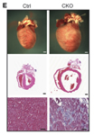
Dilated cardiomyopathy in Bag3tm1c(EUCOMM)Hmgu/Bag3tm1c(EUCOMM)Hmgu Tg(Myhca-cre)1Abel/0 mice
homeostasis/metabolism
mortality/aging
|
|
• mice are susceptible to premature death, with only 9% of mice surviving past 20 months of age
|
cardiovascular system
|
|
• cardiac enlargement in 6 month old mice
|
|
|
• heart weight to body weight and heart weight to tibia length ratios are increased at 4 months of age
|
|
|
• severe fibrosis in surviving 6 month old mice
|
|
|
• peak rate of pressure rise and peak rate of pressure decline are reduced as early as 10 weeks of age, indicating early impairment of cardiac contractile function
• single myocyte contraction induced by field stimulation is reduced
• however, the profiles of induced calcium transients, including amplitude and tau, are not affected
|
|
|
• neonatal cardiomyocytes exhibit suppression of autophagosome formation following bafilomycin-A1 and chloroquine treatment indicating impaired autophagy
|
|
|
• echocardiography shows an age-dependent decrease in left ventricular systolic function, and left ventricle chamber dilation as evidenced by an increase in end-diastolic left ventricle internal diameter and end-systolic left ventricle internal diameter
|
|
|
• single myocyte contraction induced by field stimulation is reduced
|
cellular
|
|
• neonatal cardiomyocytes exhibit suppression of autophagosome formation following bafilomycin-A1 and chloroquine treatment
|
|
|
• neonatal cardiomyocytes exhibit suppression of autophagosome formation following bafilomycin-A1 and chloroquine treatment indicating impaired autophagy
|
muscle
|
|
• peak rate of pressure rise and peak rate of pressure decline are reduced as early as 10 weeks of age, indicating early impairment of cardiac contractile function
• single myocyte contraction induced by field stimulation is reduced
• however, the profiles of induced calcium transients, including amplitude and tau, are not affected
|
growth/size/body
|
|
• cardiac enlargement in 6 month old mice
|
|
|
• heart weight to body weight and heart weight to tibia length ratios are increased at 4 months of age
|
|
|
| Find Mice |
Using the International Mouse Strain Resource (IMSR)
Mouse lines carrying:
Dot1ltm1.1Tche mutation
(0 available);
any
Dot1l mutation
(69 available)
Dot1ltm1Tche mutation
(0 available);
any
Dot1l mutation
(69 available)
Tg(Myhca-cre)1Abel mutation
(0 available)
|
|
|
mortality/aging
|
|
• mice surviving the early postnatal period died by 6 mo of age
|
|
|
• sudden death was observed in 50% of the mutant mice within 2 wk after birth
|
cardiovascular system
|
|
• gross changes in heart morphology (such as deviation from an elliptical shape to a more spherical one and increased heart mass) are observed
• RT-qPCR demonstrated that expression of the fetal genes Myh7, Acta1, Nppa, and Nppb is up-regulated in mutant hearts, and adult gene Myh6 is down-regulated indicating reactivation of a fetal gene expression program
|
|
|
• heart to body weight ratios are increased compared with that of littermate controls; body weight is not significantly altered between controls and mutant mice
|
|
|
• conscious ECHOs performed on P10 pups demonstrate that mutant mice have increased left ventricular internal dimensions and volume
|
|
|
• dilation of both heart chambers
|
|
|
• reactive fibrosis, interstitial
|
|
|
• analysis of cardiac output at P10 by measuring ejection fraction and fractional shortening reveals both are reduced by almost half in mutant mice when compared with those of control mice
|
|
|
• the percentage of proliferating cardiomyocyte cells (ratio of Ki-67-positive nuclei to total nuclei, multiplied by 100) is significantly increased in the mutant hearts compared with the control, which may contribute to the observed increase in heart mass
|
|
|
• mutant mice display at least a first-degree heart block at the atrioventricular node, with an 80% penetration of either nonsustained ventricular tachycardia, periodic third- degree heart block, or second-degree Type II heart block
|
|
|
• at P10, TUNEL staining reveals a dramatic increase in apoptotic cell death in mutant hearts compared with the control
|
cellular
|
|
• reactive fibrosis, interstitial
|
|
|
• the percentage of proliferating cardiomyocyte cells (ratio of Ki-67-positive nuclei to total nuclei, multiplied by 100) is significantly increased in the mutant hearts compared with the control, which may contribute to the observed increase in heart mass
|
|
|
• at P10, TUNEL staining reveals a dramatic increase in apoptotic cell death in mutant hearts compared with the control
|
muscle
|
|
• transmission electron microscopic (TEM) analysis reveals a significant increase of vacuoles in mutant myocytes, suggesting an increase in autophagic cell death
|
|
|
• the percentage of proliferating cardiomyocyte cells (ratio of Ki-67-positive nuclei to total nuclei, multiplied by 100) is significantly increased in the mutant hearts compared with the control, which may contribute to the observed increase in heart mass
|
|
|
• at P10, TUNEL staining reveals a dramatic increase in apoptotic cell death in mutant hearts compared with the control
|
growth/size/body
|
|
• heart to body weight ratios are increased compared with that of littermate controls; body weight is not significantly altered between controls and mutant mice
|
|
|
| Find Mice |
Using the International Mouse Strain Resource (IMSR)
Mouse lines carrying:
Mapk7tm1Jdl mutation
(0 available);
any
Mapk7 mutation
(32 available)
Tg(Myhca-cre)1Abel mutation
(0 available)
|
|
|
normal phenotype
|
|
• mice were viable and showed no overt defects through 1 year of age
|
 Analysis Tools